Advancing CT scan technology using 3D printed patient surrogates and AI
Friday, 28 July, 2023
Listen to the (opens in a new window)podcast

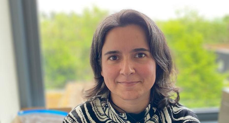
Irene Hernandez Giron (pictured) is Assistant Professor in the School of Physics at University College Dublin. She is a medical imaging researcher interested in developing what are called anthropomorphic phantoms - 3d models used as patient surrogates to test the image quality of CT scan machines. She speaks about this and about the use of artificial intelligence in medical imaging.
Legend has it thatThe Beatles’ success helped to fund one of the greatest medical advances of the last century, when their record label EMI invested in research resulting in the computed tomography (CT) machine.
Since their invention in 1971, CT scanners, which combine a series of X-ray projections taken at different angles around the body to create a volumetric image, have been a valuable medical tool to help physicians diagnose disease and injury, plan operations and monitor the effectiveness of treatments like chemotherapy.
Every year an estimated 375 million CT procedures are carried out globally - increasing by 3% to 4% annually - so it is crucial that these machines produce images of the highest possible quality.
“But the way we test medical imaging devices is still based on methods we developed 30 or 40 years ago, so they are of limited use for the new systems we have,” complains Dr Irene Hernandez Giron. “We need test objects that look more like the patient because otherwise we cannot really evaluate and optimise how the images are produced. That’s one of the driving forces to try to have more anthropomorphic phantoms.”
Anthropomorphic phantoms - or 3D models - are objects that simulate patients, made of materials with comparable tissue characteristics.
“In X-ray imaging, it's very important that we find materials that mimic the tissues we want to target in the patient.”
Dr Hernandez-Giro shows a phantom of lung vessels (pictured) - it looks like a little grey tree in a matching oval frame - she had made using 3D printed plastic.
“Plastics are not exactly like human tissue, but they need to be close enough that when I look at them in the CT image, they're going to look like, in my case, blood vessels.”
Research has already mathematically explained how lung vessels grow and she designs her lung vessel phantoms using these fractal equations.
“The advantage to having the mathematical model is that you have full control over how fast your vessels are going to change in shape. And once you design your model, you have what we call ground truth. You have completely characterised your patient.”
She then places the lung vessels phantom into the CT machine and conducts a quality assessment (QA) of the resulting X-ray images.
A little like how the Fab Four had body doubles who filmed the dangerous skiing scenes in their 1965 movie, Help!, CT scanners require patient surrogates - rather than the patients themselves - to facilitate tweaking the imaging protocols in the machines.
“When you have X-ray imaging, you have the dose to consider so it's not ethical just to irradiate the patient over and over again because there's a cancer risk. But with the anthropomorphic surrogates I can scan them as many times as I want.”
They still need to be handled and stored safely and Dr Hernandez-Giron jokes of keeping her phantoms “in a dark closet like a baby vampire at all times to protect them”, as plastics are sensitive to light and humidity.
Having joined UCD in January, she is now working on a project, with an Irish student, using phantoms of the thorax and lung vessels.
“With another student I’m looking at lesions and I'm focusing on lung nodules because there are large lung cancer screening programmes in Europe and the USA. That's why I focus a lot on thorax imaging because many patients are going to be affected by that type of imaging in the future.”
Along with trying to incorporate these potentially cancerous nodules into her lung vessel phantoms, Dr Hernandez-Giron is also trying to mimic diseases of the parenchyma - the spongy tissue that surrounds the lung vessels.
“St Vincent’s is a reference centre for Cystic Fibrosis. So it's very important, again, that we find ways to optimise those imaging protocols because it really affects the patients.”
Commercially available patient surrogates are expensive “and not everyone has €30,000 available” to buy one.
“So hopefully with my stay here I can enable the hospitals in Dublin to have access to this technology pretty quickly in the coming couple of years.”
Commercially available phantoms “normally represent only male patients” too.
“I want them to be more inclusive and that's why I focus on 3D printing because it's relatively affordable to do. Hopefully in the future we can have a variety of phantoms in each hospital to assess image quality. We should have phantoms for paediatrics, for women, for men, for people with different body types, because that really affects image quality”
Dr Hernandez-Giron is also interested in image perception by AI-based algorithms for diagnosis and image reconstruction, using 3D printed phantoms in their evaluation.
“If an algorithm tells me, ‘I've never seen these images, they don't come from a patient’, I would be okay with that. At the moment, most of those commercial algorithms always give you an answer. A clinician would not necessarily always give you an answer. Sometimes they are uncertain and they tell you that.”
She says we need Explainable AI for medical image diagnosis.
“The X-ray image workload is unsustainable for radiologists and clinicians. But we need to know the limitations of these AI tools we're going to have in place. Even if it's equivalent to the less experienced radiologists in our department, but it's consistent, it's a good tool to incorporate.”
For more information contact (opens in a new window)irene.hernandezgiron@ucd.ie
Listen to the (opens in a new window)podcast
Originally published on June 22, 2023