In a pilot study, fat deposition has been quantified as a percentage of total lung volume on chest CT published today in Respirology.
Pulmonology Fellow and PhD candidate Marissa O’Callaghan, who works in the Keane/McCarthy lab in UCD says, ‘’Our aim was to determine if lipid could be identified and measured in IPF, first on lung tissue, and then chest CT, and to assess if this correlated with disease burden.’’
The exact pathophysiology of IPF remains unknown. Treatment options are limited to drugs that slow disease progression and in some instances lung transplantation, but ultimately, mortality for this disease remains high.
There has been increasing interest in the potential role of lipids in the lung and specifically in fibrosis. Lipid is responsible for 90% of the composition of surfactant and surfactant is essential to maintain alveolar surface tension. A lack of surfactant at birth leads to respiratory failure and in some inherited surfactant disorders, fibrosis.
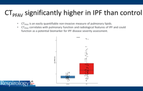
The study was designed and performed in collaboration  with University College of Los Angeles (UCLA) and National Jewish Health (NJH), Denver. Lipidomic analysis demonstrated increased lipid deposition in lung tissue from patients with a diagnosis of IPF compared to controls. The team used software to detect fat within the lung parenchyma on chest CT by measuring its attenuation volume (CTPFAV). 73 IPF and 44 control CTs were analysed.
They found that the median fat deposited within the lung parenchyma or CTPFAV was significantly increased in IPF compared to control. CTPFAV negatively correlated with lung function and in a multivariable predictive model, CTPFAV outperformed both FVC and DLCO in terms of distinguishing IPF from the control. CTPFAV also correlated with the presence of honeycombing and reticulation on chest CT but not with ground glass opacity, a widely accepted radiological marker of inflammation.
 As Dr O’Callaghan states, ‘’This is the first time that fat deposition has been quantified as a percentage of total lung volume on chest CT. Using this radiological marker (CTPFAV), we have shown an increase in lung fat in IPF compared to control. CTPFAV correlates with pulmonary function testing and radiological features of fibrosis. Measuring lung fat in this manner is non-invasive and low cost and it could function as a biomarker for disease severity in IPF.’’